MikroScan Launches Quick and Effective Real-Time Telepathology Solution

Last week, I was asked to test drive a new telepathology product from MikroScan Technologies. Their technical director, Victor Casas, explained to me very well the issues with telepathology and how MikroScan is addressing it.
He said “After a couple of years of diving head in to the world of live remote microscopy, we’ve learned some hard lessons, and have had a chance to really get an understanding of what the market needs and wants, and how to make it happen. The first and foremost condition of any telepathology device is that it must be easy to use, on both ends, for Technician and Pathologist. If it fails that test, it’s over. It must at the very basic, provide a quality image appropriate to the task set forth, and it must also have good response time, so that when the person driving it says that it’s time to move, or focus, that it does so. Initial use of the MikroScan with standard screen sharing software has been met with some success, but we’ve found a better way, through Live-Q. We’ve gotten input from Pathologists globally, have implemented many of their suggestions, and have come up with a live telepathology tool that is the kind of tool that really enables remote control microscopy across large geographical areas.”
To test it, I did what any good software tester would do, I took my Apple Laptop, grabbed a Latte, hooked up to the public wireless network at Starbucks, and signed in and called Victor, who walked me through the use of the program. To my astonishment, it was very fast and responsive, even on a shared wireless connected, the images were of very high quality, and it was really easy to use, mostly through keyboard navigation.
The technician loads up the sample and presses a button to indicate how many specimens are loaded, then walks away, their job is done for now. The scanner then makes a prescan of each slide, which takes about 20 second per slide, so in about a minute, both slides are available to be looked at through an innovative control panel, overlaid over full screen live video image.
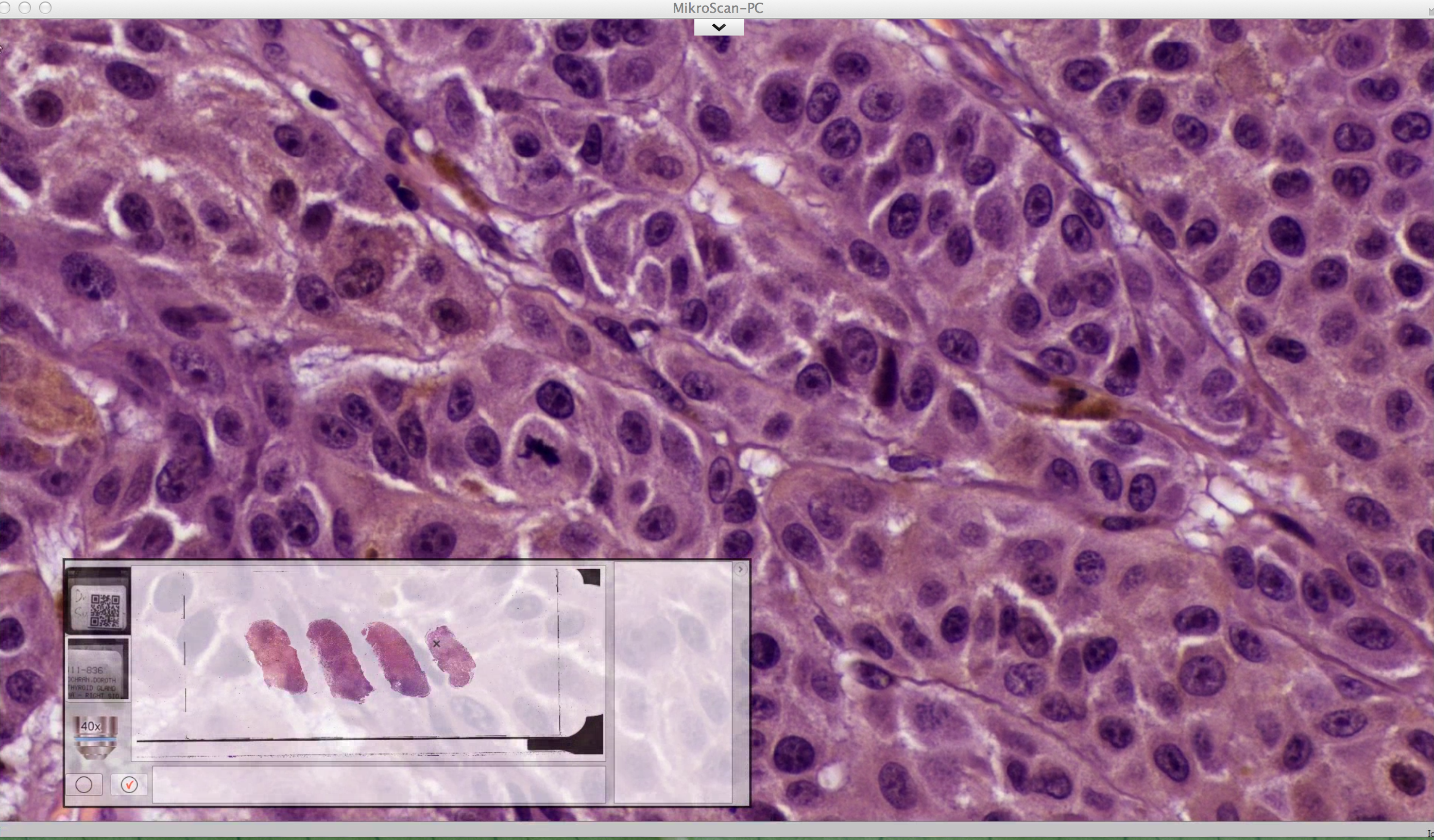
The control panel has the preview of each slide, a picture of their label, any barcode data, and a couple of extra buttons. The control panel is movable around the screen and is semi transparent so you can see what’s going on behind it, and is also minimizable so you can just view full screen video if you’d like. The buttons allow you to make “virtual inkmarks” on the slide or slides, mark them on the prescan and return to them later with the push of a button. It lets you with the push of one button, save a high resolution image (1920×1080) of the current field, as many as you’d like. It also keeps track of where you’ve viewed. When you save the case, you save the prescan images, the label images, the hotmap history track, and any virtual inkmarks, as well as any images you’ve saved into a folder defined by either barcode data that was initially scanned, or what you’ve named the specimen, which can be saved to a local hard drive, a network drive or wherever you would like. When you’re done, and go home, an alarm is sounded in the technician’s lab indicating that it’s time to change the slide, or you’re done with the case.
In summary, this system works on every level. It’s fast, it’s responsive, it’s intuitive, it’s high quality, and it’s easy to use, and delivers everything it promises. With it’s ability to scan small volumes of slides, and a real-world remote microscopy solution, at the small price tag of the MikroScan, there’s no reason it’s not in multiples in pathology departments for quick frozen sections, peer to peer review, immediate FNAs and proforma QA cases.


