ZEISS Axio Scan.Z1 Advanced Digital Imaging System Improves Pathology Research

![]() At the University of Pittsburgh’s Department of Transplant Pathology, scientists use advanced digital imaging systems to enhance their research into the immunobiological mechanisms of tolerance and chronic rejection in solid organ transplantation.
At the University of Pittsburgh’s Department of Transplant Pathology, scientists use advanced digital imaging systems to enhance their research into the immunobiological mechanisms of tolerance and chronic rejection in solid organ transplantation.
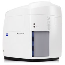
 A pivotal innovative tool is the ZEISS Axio Scan.Z1, which is an automated microscope developed by Carl Zeiss Microscopy. This tool enables researchers to convert fixed tissue sections and cytologic specimens using bright-field and fluorescence illumination into digital image files that can be viewed and analyzed on a computer screen instead of being tethered to the microscope. This approach gives researchers “next generation pathology” capabilities, including functionality to compare images side by side, software algorithms for counting, comparison, analysis, and quantification of specific tissue areas or cells of interest to greatly expand the type and amount of data that can be harvested from a tissue biopsy.
Twenty years of digital imaging advancements
The University of Pittsburgh’s Department of Pathology was an early adopter of digital imaging systems, beginning in the late 1990s. The department, which includes specific divisions for transplantation and neuropathology, used the technology to support its clinical operations by offering second opinion capability and educational imaging resources to other practitioners, as well to enhance its research by facilitating image quantification.
 According to Andrew Lesniak, Senior Architect for the department’s Pathology Informatics Group, the department entered into a collaborative relationship with Carl Zeiss Microscopy in 2007, when it began using the ZEISS MIRAX line of scanners to enhance its routine transmitted light scanning and to use fluorescence (multiplexing) to study tolerance induction in the liver, kidney and heart. The ZEISS MIRAX was known at the time for having the best resolution in the industry, and offered the highest level of Fluorescence acquisition functionality. The department is now transitioning over to the ZEISS Axio Scan.Z1 as its main imaging platform for whole slide imaging (WSI) acquisition.
Lesniak and his colleagues believe that the ZEISS Axio Scan.Z1 offers the best optical and imaging hardware, coupled with innovative visualization and analysis software, to maximize the information that can be obtained from a tissue image. He cites the fact that ZEISS actively engages in the scientific process to understand and help solve the biological problems being studied as the reason the relationship transitioned from a customer to a collaborator.
“ZEISS does not stop at building the tool; they work with investigators to ensure you realize the range of capabilities the tool provides in solving biological problems”.
Working closely with ZEISS, the department made suggestions in terms of features and functionality, adapting and tweaking the technology to maximize the value that can be achieved from scanning a biopsy. They funneled this product level feedback through the product development group, and the input was ultimately used in the development of the ZEISS Axio Scan.Z1.
“What sets ZEISS apart is that they understand some of the most challenging aspects of the image acquisition process and develop their technology to solve these problems.”
Under the hood of ZEISS Axio Scan.Z1
The ZEISS Axio Scan.Z1 is an automated microscope with an innovative tray design that captures the entire specimen area of a microscope slide, including the edge. The self-calibrating automated slide scanner presents specimens on high-quality virtual slides just minutes later. For fluorescence applications, filter wheels switch wavelengths in just 50 milliseconds.
Sensitive cameras and maximally corrected optics achieve optimal image quality. For fluorescence acquisition, maximum protection against photobleaching (fading) of the sample is ensured by a ZEISS Colibri.2 UV-free LED light source, as well as a focus finder with oblique illumination, called the ring aperture contrast.
The instrument is operated by proprietary ZEISS ZEN imaging software, which allows users to work with pre-defined recording parameters automatically or select all settings individually. The user interface is specially adapted to research workflow. The University of Pittsburgh researchers can decide at the outset how many microscope slides and which detection modes and camera they need for a particular experiment or seeting. The system is designed as a platform technology, with ability to upgrade the ZEISS Axio Scan.Z1 as their tasks expand.
The virtual slides can be organized in the ZEISS ZEN Browser database. This software presents data online and is cross-platform compatible via an intuitive web interface that keeps images filed together with metadata and supporting documents. With ZEISS ZEN browser, users can also insert annotations and view images in transmitted light and with full multi-channel fluorescence controls. The free ZEISS ZEN browser mobile app for iPad and iPhone allows users to access, view, and share their images and data with colleagues independent of the operating system – even when traveling.
Microscope enhances critical transplantation research
The Department of Transplant Pathology’s research focuses on innovating histopathological (microscopic tissue examination) multiplexing, imaging and analysis techniques to capture and quantify the spatial and temporal data that is present in a digital whole slide scan. The university’s investigation of the benefits of whole slide imaging technology is being performed as part of large scale research studies conducted under in collaboration with NIH-sponsored Clinical Trials in Organ Transplantation and the Immune Tolerance Network, an international consortium of clinical and basic investigators dedicated to accelerating the clinical development of immune tolerance therapies in solid organ transplantation. Tissue biopsies are flown to the university from around the U.S. and are examined digitally and analyzed with multiplex-enabled imaging systems.
Lesniak explains that focusing is a challenge when dealing with whole slide imaging in fluorescence, and the new ZEISS Axio Scan.Z1 functionality provides a flexible blend of hardware and software functionality to make capturing sensitive specimens possible. ZEISS Axio Scan.Z1’s increased resolution improves human and software-assisted analysis and interpretation leading to novel insights into tissue biology that have not been routinely possible prior to implementation of these tools.
The data they capture from the ZEISS Axio Scan.Z1 provides a more in-depth, quantitative, and mechanistic view of the complex relationships present among cells in the tissues. For example the number, density, and proximity of specifically defined cell types to other cell types or tissue structures can be determined.. The information can be linked to other data sources for cross-platform analyses that can link tissue events to serum markers and to various genomics analyses.
“This is crossplatform translational research,” says Lesniak. “We see in the Axio Scan.Z1 the capability to perform high sensitivity imaging, as well as increase our lab’s output by using the batch scanning capabilities for larger datasets with confidence of a good image product.”
Source: ZEISS


