Uncover a Masterpiece at the American Association for Immunology (AAI) — Pittsburgh, PA – May 2-6

Visit the PerkinElmer booth (#215) at the AAI Annual Meeting and learn about our full range of quantitative pathology imaging solutions. Label, image and count multiple phenotypes of immune cells in situ in FFPE tissue. Our technology platform and biomarker reagentsenable you to study the same sorts of complex phenotypes you currently study in flow cytometry without compromising the morphology of the tumor – the analysis takes place in situ in the tumor microenvironment.
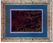
Stop by our booth and register to win an IHC work of Art.
Our solutions bring together several different technologies for measuring immune cells in situ, our Vectra Automated Multispectral Imagingsystem, the Opal™ Multiplex Tissue Staining workflow using covalent labelling with tyramide signal amplification (TSA™) reagents and our inForm® Tissue Finder™ quantitative pathology analysis software.
Application Benefits
- Automatically label, measure, visualize, count and compare multiple immune cell phenotypes within the tumor microenvironment
- Mutiplex up to 7 markers simultaneously in a single section using any secondary antibody species combination in fluorescence and up to 3 in brightfield
- Image and separate multiple overlapping chromogenic or fluorescent markers to view individual expression patterns
- Train the system to automatically detect tumor regions (i.e. invading edges, margins, etc) in whole tissue sections
- Perform per-cell quantitative analysis per marker within biologically relevant morphologies, e.g., tumor and stromal tissue compartments


