Apparently it is official…pathologists have been replaced by Google. Just like those folding maps, the yellow pages, the white pages, newspapers, racing forms, microfiche, books and other annoying analog nuances of daily life.
The original story is of course not “just”, unless “just” means news going back a month as shown on CNN: Google uses AI to help diagnose breast cancer. In my post earlier this week: A.I. Versus M.D. – Either Way – Pathologists Do Not Diagnose Cancer – Part 1 I hoped to be retired before imaging robots take over our paneled offices and replace us and our microscopes but according to The Motley Fool, us human pathologists have already been beat trying to get the right diagnosis in a timely, cost-efficient manner.
While the original Google blog story and subsequent articles and interviews on the matter cite that “Pathologists are responsible for reviewing all the biological tissues visible on a slide. However, there can be many slides per patient, each of which is 10+ gigapixels when digitized at 40X magnification. Imagine having to go through a thousand 10 megapixel (MP) photos, and having to be responsible for every pixel. Needless to say, this is a lot of data to cover, and often time is limited.” The Motley Fool provides what they mention as “Radiology technician reviewing mammography results” and “Woman reviewing X-Rays on lighted panel” for their photo captions in their article.
Google points out as well that these technologies will allow pathologists to work more efficiently, find the needle in the haystack type approach, but it will still be up to the pathologist for the final determination. Not what The Motley Fool claims however though, instead, not only do the stock images that are in this article show radiologists, or radiology technicians examining radiology studies, but one of them is looking at head CT images printed on pieces of plastic on something called a lighted panel!
Seriously Fool.com?
In the follow up piece to this when thousands of pathologists are unemployed forced to go back into primary care or construction or blogging full time, can you at least include a picture of a pathologist with a glass slide and a microscope or a lab coat or Erlenmeyer flask with a blue liquid in it or something other than a head CT examination on plastic on a lighted panel when writing about AI in pathology for cancer diagnoses?!?
From Fool.com:
Artificial intelligence is tailor-made for sifting through massive amounts of data to find patterns. GoogLeNet AI just used image recognition and bested human doctors at detecting breast cancer.
The science of deep learning, a sub-discipline of artificial intelligence (AI), is only a recent development in the grand scheme of things, but during its short existence, it has been producing some impressive technological achievements. Advances in image recognition, language understanding, and translation have led to the development of virtual assistants, smart home speakers, and gains in cybersecurity, and they are leading the charge toward autonomous driving. Now, companies have found a way to use those AI smarts to fight cancer.Deep learning involves the construction of artificial neural networks, using software and complex algorithms to recreate the capacity of the human brain to learn. These learning computers have a particular knack for sifting through vast amounts of data and recognizing patterns, getting smarter as they go. The first breakthrough involved feeding a system thousands of pictures of cats until the program was able to recognize a cat on its own.
GoogLeNet AI provides groundbreaking cancer research! Image source: Getty Images. Alternative text: Radiology technician reviewing mammography results.
GoogLeNet AI provides groundbreaking cancer research! Image source: Getty Images.
Recognizing breast cancer tumors
This ability to identify patterns has led to a significant breakthrough in the area of breast cancer research. Last month, in a paper titled Detecting Cancer Metastases on Gigapixel Pathology Images, Alphabet Inc. (NASDAQ:GOOG) (NASDAQ:GOOGL) division Google announced that it had created a neural network that could analyze medical images and identify tumors with a greater degree of accuracy than human pathologists. The study revealed that the company, using its GoogLeNet AI, reviewed thousands of medical images supplied by a Dutch university and was able to identify malignant tumors in breast cancer images with an 89% accuracy rate, compared to 73% for its human counterparts. In a blog, Google researchers explained:
Pathologists are responsible for reviewing all the biological tissues visible on a slide. However, there can be many slides per patient, each of which is 10+ gigapixels when digitized at 40X magnification. Imagine having to go through a thousand 10 megapixel (MP) photos, and having to be responsible for every pixel. Needless to say, this is a lot of data to cover, and often time is limited.
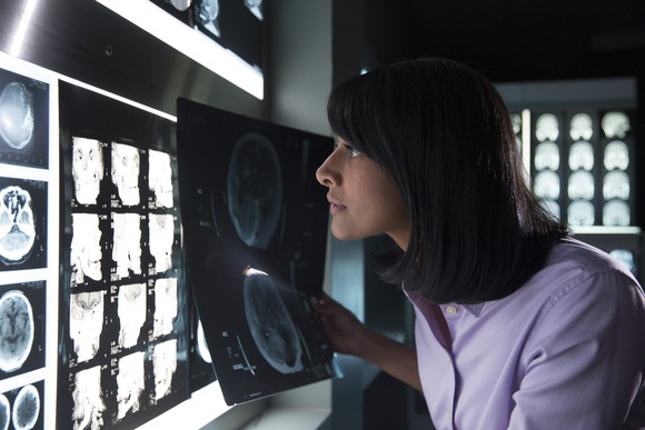
IBM enhances Watson’s ability to “see” medical images. Image source: IBM. Alternative text: Woman reviewing X-Rays on lighted panel.
This technology has the potential to provide initial screenings, allowing doctors to review only those images that have been flagged as potentially cancerous. The system still requires improvement, as it generated a number of false positives — identifying cancerous cells where none were present. So, while AI won’t be replacing pathologists anytime soon, these algorithms could be used to pre-screen images and not only reduce the workload on doctors, but also serve parts of the world where pathologists are in short supply.
Read full article on Fool.com here.