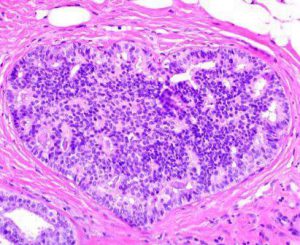
Whole Slide Images as Good as Glass for Breast Biopsies
 A recent publication in the Journal of Pathology Informatics a group of researchers from the University of Miami using a BioImagene scanner report on their experience with review of breast biopsies with whole slide images as compared with conventional microscopy. As we are increasingly seeing, the intraobserver variability in diagnosis of the core needle biopsies of the breast, in this use case, by whole slide imaging was the same as conventional glass slide microscopy. In other words, the same pathologist looking at the same slide, with a washout period using digital pathology versus conventional light microscopy did not yield a difference in diagnosis or intraobserver variability. Difficult cases, it turns out, are difficult either way as one might predict — a challenging case is a challenging case is challenging case — depending on day of week, time of day, one might disagree with his or herself when they review the same slide in another place or at another date and time. Subtle differences in usual ductal hyperplasia and atypical ductal hyperplasia can be very subjective among experts (inter observer) and among single readers (intraobserver). Encourage you to check out the paper below and their discussion and commentary for a nice review of the growing body of work within each respective sub-specialty field of surgical pathology trying to answer the question “Is whole slide imaging inferior to the light microscope” and discovering that whole slide imaging is as good, if not better than the tried and true light microscope (using this as the “gold standard” diagnosis).
Reyes C, Ikpatt OF, Nadji M, Cote RJ. Intra-observer reproducibility of whole slide imaging for the primary diagnosis of breast needle biopsies. J Pathol Inform [serial online] 2014 [cited 2014 Mar 2];5:5. Available from: http://www.jpathinformatics.org/text.asp?2014/5/1/5/127814
Abstract
Background: Automated whole slide imaging (WSI), also known as virtual microscopy is rapidly becoming an important tool in diagnostic pathology. Currently, the primary utilization of the technique is for transmission of digital images, for second opinion consultation, as well as for quality assurance and education. The high-resolution of digital images along with the refinement of technology could now allow for WSI to be used as an alternative to conventional microscopy (CM) as a first line diagnostic platform. However, the accuracy and reproducibility of the technology for the routine histopathologic diagnosis has not been established yet. This study was undertaken to compare the intra-observer variability of WSI and CM in the primary diagnosis of breast biopsies. Materials and Methods: One hundred and three consecutive core needle biopsies of breast were selected for this study. Each slide was digitally scanned and the images were stored in a shared file. Three board-certified pathologists independently reviewed the glass slides by CM first, and in an interval of 2-3 weeks for the 2nd time to establish their baseline CM versus CM reproducibility. They then reviewed the digital images of all cases following the same interval of time to compare the reproducibility of WSI versus CM for each observer. The diagnostic categories included the typical range of benign and malignant mammary lesions. Results: The intra-observer variability for CM versus CM was 4%, 7%, and 0% for observers 1, 2, and 3 respectively. The diagnostic variability for WSI versus CM was 1%, 4%, and 1% for the same observers. All diagnostic disagreements were between ductal hyperplasia and atypical ductal hyperplasia. There was no intra-observer disagreement in the diagnosis of benign versus malignant disease. Conclusions: The intra-observer variability in the diagnosis of the core needle biopsies of the breast by high-resolution, WSI was the same as conventional glass slide microscopy. These results suggest that, WSI could be used similar to CM for the initial diagnosis of breast biopsies.
Reyes C, Ikpatt OF, Nadji M, Cote RJ. Intra-observer reproducibility of whole slide imaging for the primary diagnosis of breast needle biopsies. J Pathol Inform [serial online] 2014 [cited 2014 Mar 2];5:5. Available from: http://www.jpathinformatics.org/text.asp?2014/5/1/5/127814


