Quantify Biomarkers in situ in Tissue Sections with PerkinElmer’s NEW inForm 2.1 Tissue Finder software

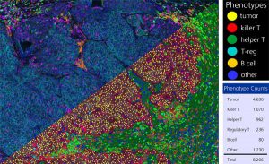
Breast cancer tissue showing presence of helper and cytotoxic T-cells in and out of tumor and stroma using inForm software. Upper left: Umixed composite image with cytokeratin (cyan), killer T-cells (purple), helper T-cells (green), B-cells (red), PD-L1 (orange), FOXP3 (yellow). Bottom right: Segmented cell and tissue phenotypes and counts. Upper right: Color key for phenotyped cells. Lower right: Cell count by phenotype. Work performed in collaboration with Dr. Beth Mittendorf at MD Anderson.
Extracting meaningful information from intact tissue is becoming ever more critical as researchers look for better ways to characterize molecular activity of cells within their native micro-environments. With PerkinElmer’s new inForm® 2.1 Tissue Finder™ advanced image analysis software you can accurately quantify, visualize and phenotype cells in situ in tissue sections. inForm Tissue Finder automates the detection and segmentation of specific tissue types using patented user-trainable algorithms to recognize morphological patterns and spectral signatures. The new software also includes Pathology Views™ for visualization of multi-label fluorescence imagery in traditional brightfield mode (as if you were looking at conventional H&E, DAB and hematoxylin images).
Features of inForm 2.1
- Visualizes, analyzes, quantifies and phenotypes immune and other cells in situ in solid tissue
- Separates weakly expressing and overlapping markers
- Cellular analysis of H&E, IHC and immunofluorescence in FFPE tissues sections
- Automatic identification of specific cell and tissue compartment types using trainable feature-recognition algorithms
- Pathology Views displays fluorescence imagery as traditional H&E, DAB and hematoxylin

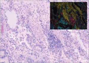
Pathology View rendering of original fluorescence image, seen as if it were in standard brightfield mode, providing a familiar view for a pathologist used to seeing H&E, DAB and hematoxylin.
Learn more about our new inForm 2.1 analysis software.
Visit PerkinElmer at the following conferences to learn more about our new inForm 2.1 image analysis software and Mantra® Workstation for multiplex biomarker analysis or visit our website for more information.
AACR – Apr 18-22, Philadelphia, PA
AAI – May 8-12, New Orleans, LA
CIMT Cancer Immunotherapy – May 11-13, Mainz, Germany
International Symposium on Immunotherapy – May 15-16, London
ASCO – May 29 – Jun 2, Chicago, IL


