November 26, 2019
Tissue Classification

![]() Marie-Louise Loupart, Digital Pathology Software Specialist – Leica Biosystems
Marie-Louise Loupart, Digital Pathology Software Specialist – Leica Biosystems
- Today, we are surrounded by Artificial Intelligence (AI) and often don’t even know it’s there. AI systems make headlines when something goes wrong – driverless cars appear to be our current main stream pre-occupation with the dangers of AI.
- AI is making inroads into medical practice but it is slow progress as it seems we prefer our experts to be human when it comes to our health. AI in the field of Pathology is trailing behind Radiography but there is much focus on getting AI into day-to-day pathology. What can it do for us here?
- The honest answer is that no one knows! We can theorize, make extrapolations, and endeavor not to crash this particular car…
- Extrapolate from what? When we talk about AI in Pathology we’re really talking about Deep Learning… and the forerunner of that is Machine Learning (actually DL is a subset of ML, that in turn is a subset of AI, for those individuals who are most particular in their terminology).
- So, is it reasonable to use ML as the jump off point? After all, no ML system has been approved as a medical device for Pathology. Is ML ‘any good’? That depends on what you want it for.
- As an Image Analysis Specialist at Leica Biosystems, I’ve been working with Aperio GENIE for 3 years – for customer demos, proofs of concept and ultimately trying to generate a useful tool in a real lab. I’ve learnt that the pathology lab is a very different reality…
- Aperio GENIE algorithm has been a commercial product for over a decade, after Aperio, Inc. was granted license to develop it for use on pathology images. The algorithm’s original incarnation was to interpret geo-satellite imagery by Los Alamos National Laboratories.
- From geography to cancer is not such a large step when you look at typical examples of each and maybe squint a bit…
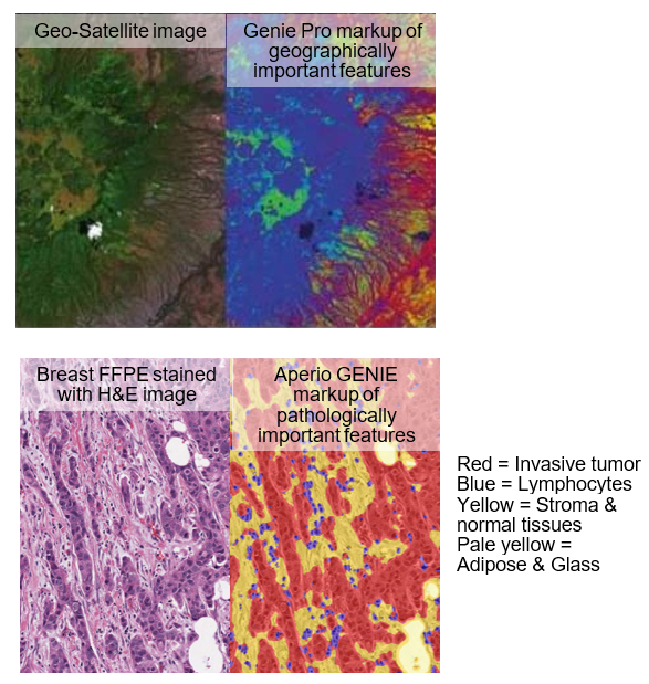
- Machine Learning classifiers are very specific – they can be robust when trained on a cohort of slides that were produced in the same laboratory production line, providing batch-independent tissue classification. Rarely can these classifiers be shared between multiple sites – they are LOCAL classifiers.
- What sends this car off the road? Variation in tissue processing, stain reagents and methods, and scanning/imaging, all contribute variability in the final digital image. Lock down as many variables as possible and the dataset becomes easy to generate for a robust classifier. The peer-reviewed publications by Aperio GENIE users out there prove it.
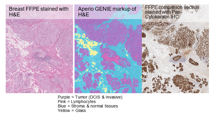
- Verifying the accuracy of any tissue classifier with comparison to expert opinion, as well as companion IHC slide, can be challenging. The lesson is ‘beware the non-adjacent section’:
- Aperio GENIE can be a liberator in the research laboratory:
- A robust classifier can act as a pre-screener for whole batches of slides – to find those with the ROIs for the expert to focus on
- No more laborious manual annotation of ROIs for analysis as the classifier will highlight the ROIs generally to a resolution of 2-5 cells
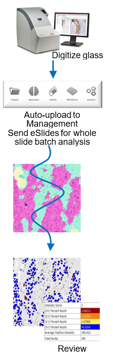
- Technicians can send images for whole slide analysis so Pathologists only need to interact with the images after the whole slide classification analysis
- It can provide non-experts with their own specific ‘expert on demand’, freeing Pathologists from regular demands on his/her time.
- Machine Learning has proved to be a great tool for research but it has 2 important weaknesses that preclude its use in the clinic. The clinic demands a higher degree of accuracy and a GLOBAL classifier. Leica Biosystems’ in-house R&D project on Deep Learning indicates it could/should provide the specificity for the ultimate clinical ‘plug-and-play’ solution.
Source: Leica Biosystems Imaging, Inc.


© 2023 Tissuepathology.com. All rights reserved. Millennial Consulting. | Web Design by Zealth Digital Marketing