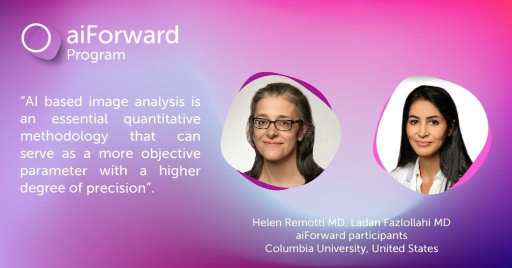
Columbia University Pathologists kick off AI powered liver research project

>Interview with pathologists Helen Remotti MD and Ladan Fazlollahi M
What do you do and what is your research focus?
We are academic pathologists with broad research interests involving liver/gastrointestinal/pancreatic benign and neoplastic diseases. Fatty liver disease has become an important area to study as it has become the leading cause of liver disease in United States and worldwide, and among the top reasons for liver transplantation. Here at Columbia University Medical Center, we receive a large number of liver biopsies from patients with alcoholic and non-alcoholic fatty liver disease. Therefore, we do have the resources and the opportunity to advance our study methods and understanding of this disease.
Why did you apply to the aiForward program?
As pathologists, we quantitate the degree of fat content in our liver biopsies as mild (5-33%), moderate (>33-66%) and severe (>66%) by estimating the percentage of fat under microscope. We also use the routine H&E slide and a trichrome-stained slide to assess the stage of fibrosis (Stage 1 to 4). There is some degree of inter-observer variation in estimating the percent fat and fibrosis stage. The data derived from this type of pathology analysis is used to direct patient care and even enrollment in clinical trials. Now with AI-based imaging techniques available, we are interested to see if these techniques could help us better estimate the fat content, differentiate the small fat-droplets from large fat-droplets and fibrosis stage in liver biopsies. In addition, AI based imaging can be used for quantitating other biomarkers.
Had you thought much about using AI in your research before applying to aiForward?
Yes. we have previously worked on multiple collaborative projects focused on pancreatic, liver, and colorectal carcinomas. We used AI-based image analysis techniques to evaluate and compare the immune cell density in tumor and stroma. I believe there is a great need for AI-based image analysis techniques in pathology research, and eventually clinical diagnosis, as we need to move from subjective analysis of tissue components to more robust and objective quantitate methods.
Tell us a little bit about your aiForward project.
We have a cohort of liver biopsies from patients with non-alcoholic steatohepatitis (NASH) and a cohort of liver biopsies from control patients (liver donors) with correlative Fibroscan liver stiffness measurements. FibroScan is a non-invasive ultrasound-based imaging technique used by hepatologists to indirectly assess the fat content and elasticity (corresponding to fibrosis) of the liver parenchyma. The aim of our study is to use the AI-image analysis platform developed by Aiforia for quantitative evaluation of fat and fibrosis in liver biopsies, and to correlate these findings with parameters obtained by routine pathology assessment and FibroScan.
What are your expectations for the program?
We would like to have quantitative data on percentage of fat and fibrosis in our cohort of liver biopsies within a short timeframe (less than 6 months). Quantitation of additional biomarkers is planned for future studies.
____________
Applications to the aiForward program are accepted on a continuous basis. Find out more or apply here: https://www.aiforward.org/apply/
Find out more about Aiforia: https://www.aiforia.com/aiforia-create/


