Came across this at another blog – The Medical Quack through the Medicine 2.0 blog carnival.
Both of these blogs have some excellent content. The original story was on technologyreview.com last month with its own great items!
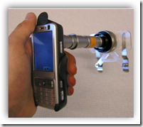
Researchers at the University of California, Berkeley, have developed a modular, high-magnification microscope attachment for cell phones. The device will enable health workers in remote, rural areas to take high-resolution images of a patient’s blood cells using a cell-phone camera, and then transmit the photos to experts at medical centers.

The total cost of the first prototype, built from off-the-shelf components, was $75. The current version provides its own sample illumination from cheap, low-power LEDs. The device comes in two versions: with a magnification of about 5 times, for taking images of moles and rashes, and with a magnification of about 60 times, for capturing the details of blood cells and parasites.
The scheme is to train local personnel and provide them with the necessary equipment to take pictures of patients’ blood on special slides, and then phone in the images to specialists who can identify and count malaria parasites.
————————————————————————————————————–
My comments:
While the device for $75 is very interesting and allows for inexpensive imaging of clinical findings and microscope images, the concept has been around in dermatology and the clinical laboratory for years. I have a previous posting about resolution with a low-resolution image (by todays standards) of malaria taken through a even lower resolution microscope objective. This was from 2002. It was taken by an infectious disease fellow for the next days’ morning conference. It could have just as easily been used for consultation. Is this P. falciparum? on a short e-mail or text message with image attached. At the same institution dermatology was doing the same between primary care providers "in the field" and dermatology "at the main hospital". Cameras were provided, web-enabled secure system stood up to upload, download and track who did what when, from where and who saw it when and where, what he/she said and ultimately patient disposition for this encounter.
Ultimately, these types of consultations (posted as "schemes" above) fail becuase of lack of clinical champion, technophobia on part of consulting physician or an answer of "Yes, that looks like an atypical nevus, we should see the patient ", "You are right, that is a rash, I am not sure based on your history what it is", or "We really need to see the slide to make that diagnosis". So referring physicians and pathologists go through this half a dozen times or so and think, I will just send the patient or slides and avoid these extra steps if ultimately no triage is going to occur (outside of referral which in some cases probably isn’t warranted but remote diagnosis is out of the comfort level of consultant and they are unwilling to be associated with this without laying hands on the patient or slide themselves….).
© 2023 Tissuepathology.com. All rights reserved. Millennial Consulting. | Web Design by Zealth Digital Marketing


