November 22, 2019
Tissue Classification To Direct Image Analysis of Fluorescent Images
 Marie-Louise Loupart, Digital Pathology Software Specialist
- Previously, we have examined uses for pattern recognition in brightfield images. Let us focus on what it can do for fluorescent applications.
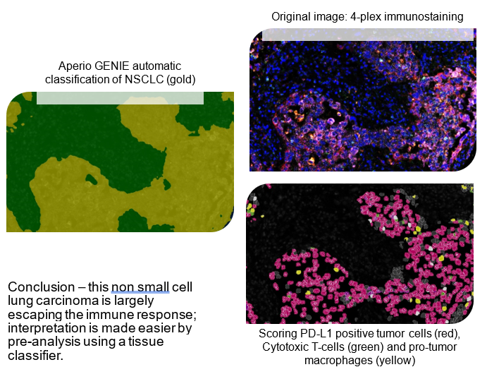
- Machine Learning classifiers are suitable for multiplex assays, ensuring the user is looking at the desired phenotypes in the correct tissue types. If the study is focused on oncology, there is no point in chasing down an immune response caused by inflammation from a prior biopsy or even an infection.

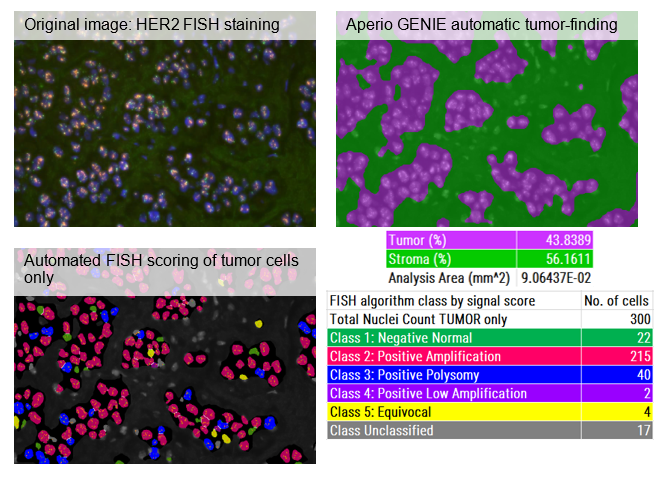
- FISH scoring of solid tumors is hampered by the lack of morphology beyond nuclear shape (DAPI) to distinguish tumor from normal. Overlapping nuclei and irregular shapes compound the issue, driving up time to score because additional fields of view must be reviewed. Aperio GENIE classification of the FISH slide will enrich the FISH scoring algorithm with tumor cells, generating not just the minimum 100-300 total cells, but across the whole slide without the need to manually annotate.
- Close scrutiny of the FISH image shows variable background intensity of the orange and green fluors. This is not uncommon and both the Aperio GENIE classifier and the FISH scoring algorithm will allow for this so you will get your FISH signals in your tumor cells scored automatically.

- Would I go back to the days of 4-color FISH and IF scoring direct from a microscope, attempting manual analysis with a clicker and pen and paper? No thanks! It was too hard to keep it all straight in my poor head and seeing what I noted down on the sheet. Digital is for me.
- In conclusion, this short series on use of pattern recognition to aid interpretation of histopathology images highlights how invaluable Machine Learning can be as a tool in Histopathology, paving the way for Deep Learning. For many years to come, both will have a specific job to do: rapid-build local Machine Learning classifiers and slow-build global Deep Learning classifiers. Who will win out in the end? Actually we will, the users of these incredible tools as they prove to us that we can rely on them to ease and deepen our knowledge and capabilities in the Digital Era of Histopathology.
Source: Leica Biosystems
© 2023 Tissuepathology.com. All rights reserved. Millennial Consulting. | Web Design by Zealth Digital Marketing


