Join this thought-provoking webinar with renowned researchers and company leaders


It’s all about location:
Novel prognostic models derived from spatial analysis of the tumor-immune interface
29 July 2020 | 16.00 (London), 11:00 (New York), 08:00 (San Francisco)
Duration: 75 minutes
Biological interactions between functionally distinct immune and tumor cell populations can either promote or inhibit tumor growth. In this 75-minute webinar, we will hear from two research teams who have identified novel prognostic signatures using different spatial analysis and machine learning approaches to measure tumor-immune cell interactions within the tumor immune microenvironment (TIME). A short presentation on analysis tools for multiplexing, spatial analysis and deep learning will be made by Indica Labs, our webinar sponsor.
Do join this thought-provoking digital session with renowned researchers and company leaders!
![]()

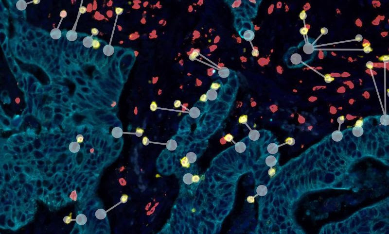
International validation of a novel spatial immuno-oncology prognostic model in stage II colorectal cancer
The tumour microenvironment (TME) plays a major role in tumour progression and patient survival outcome. Several components of this complex TME have shown to be promising prognostic factors in stage II colorectal cancer (CRC) however, they are traditionally reported in isolation of each other. In this study, we evaluated the densities and interactions of tumour infiltrating lymphocytes, macrophages and tumour buds (TBs) in order to create a more personalised prognosis for patients with stage II CRC. Multiplexed immunofluorescence and automated image analysis were used for the quantification of CD3+, CD8+ T cells, CD68+, CD163+ macrophages and TBs, across 2 sequential whole slide images. This was performed across 2 independent cohorts (training cohort: n = 113, validation cohort: n = 117). Exported features were processed through machine learning algorithms for the development of a new prognostic risk model. This model combined lymphocyte infiltration, CD68+ /CD163+ macrophage ratio and the spatial proximity of lymphocytes to TBs. When applied to the training cohort, the model stratified a patient subpopulation with 100% survival over 5 years. This result was confirmed in an independent validation cohort. The digital pathology and machine learning-based methodology reported here demonstrate the ability to identify stage II CRC patients at low risk of disease progression and who would therefore not require detailed follow-up or further unnecessary invasive or chemotherapeutic treatment.

Assessment of Intratumor Heterogeneity and Tumor/Host Interaction using Spatial Analytics
Assessment of tumor tissue biomarkers can and should go beyond routine quantification to represent an average level of expression in the sample. High-capacity digital image analysis enables various methods of spatial statistics to extract subvisual features of biomarker expression taking into account intratumour heterogeneity and tumor microenvironment context. Both aspects are crucial for the development of robust prognostic and predictive tissue biomarkers. In this presentation, we will discuss image analytics, based on hexagonal grid subsampling of image analysis data and explicit mathematical modeling of the indicators. In particular, we will focus on the Interface Zone Immunogradient indicators1 which represent tumor infiltrating lymphocyte density profiles across the automatically defined tumour/host frontline. Independent prognostic value of the indicators has been demonstrated in breast and colorectal cancer patient cohorts.


![]()