Mass Spectrometry Imaging Used For Tissue Analysis

Scientists from Imperial College London (ICL) have developed a method that uses mass spectrometry imaging (MSI) to analyse biological samples based on their chemical makeup.
The MSI technique uses technologies that reveal how hundreds or thousands of chemical components are distributed in a tissue sample.
Until now there has not been a formula to apply this technology to identify tissue types, but the ICL researchers have this week outlined a method for processing MSI data and building a database of tissue types.
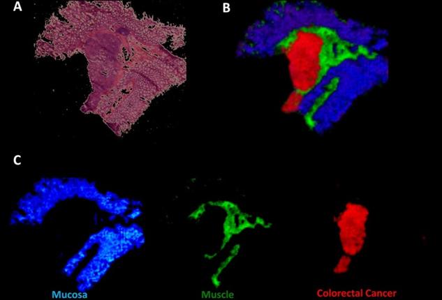
In MSI, a beam moves across the surface of a sample, producing a pixelated image. Each pixel contains data on thousands of chemicals present in that part of the sample.
By analysing numerous samples and comparing them to the results of traditional histological analysis, a computer can learn to identify different types of tissue.
A single MSI test, which takes a few hours, can provide more detailed information than standard histological tests, for example showing not just if a tissue is cancerous, what the type and sub-type of cancer, which can be important for choosing the best treatment.
ICL researcher Zoltan Takats said: “This technology can change the fundamental paradigm of histology. Instead of defining tissue types by their structure, we can define them by their chemical composition.
“This method is independent of the user – it’s based on numerical data, rather than a specialist’s eyes – and it can tell you much more in one test than histology can show in many tests.”
The team have said the discovery could also be useful in drug discovery as MSI would allow researchers to look for a new drug absorbed in the body, without using radioactive labels – the technique a pharmaceutical scientist uses to locate new drug molecules within test subjects.
In this week’s Proceedings of the National Academy of Sciences (“Chemo-informatic strategy for imaging mass spectrometry-based hyperspectral profiling of lipid signatures in colorectal cancer”), researchers at Imperial College London have outlined a recipe for processing MSI data and building a database of tissue types.
Read more: http://www.nanowerk.com/news2/biotech/newsid=33895.php#ixzz2qLrRduA5