Join PerkinElmer at the 26th European Congress of Pathology in London– August 30 – September 3, 2014

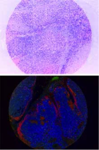 Visit our booth (#37) at ECP to learn how our quantitative pathology solutions can help analyze complex relationships among tumor and immune cells in the tumor micro-environment. Our imaging systems and reagent solutions can deliver per-cell quantitative protein expression from up to 8 markers, giving data similar to that obtained from flow cytometry, but from FFPE tissue sections. See how the Opal™ multimarker staining, imaging and analysis methodology can be used to better understand the role of multiple types of immune cells in and around the a tumor and improve our understanding of new cancer immunotherapy treatments.
Our multispectral and whole slide imaging systems are ideal for many areas of pathology research. Come see our Lamina™ multilabel slide scanner which delivers clear, high-resolution images from whole slides and Tissue Microarrays (TMAs). With Brightfield, Fluorescence and Autofluorescence Reduction modes all in one instrument, Lamina is a flexible platform for your research pathology lab.
Posters and Presentations at ECP
Poster
“Autofluorescence reduction and cross-talk correction in a whole-slide digital pathology system”
James Mansfield, Director of Quantitative Pathology Applications, PerkinElmer
Presentations
“Investigating the prognostic significance of Tumour Infiltrating Lymphocytes (TILs) in Post-Transplant Lymphoproliferative Disorder (PTLD): A novel approach”
Christian Slater, University of Manchester
“Multispectral imaging and image analysis software can accurately quantify immunophenotypically distinct Tumour Infiltrating Lymphocytes (TIL) using chromogenic multiplexed immunohistochemistry (IHC) in lymphoid Tissue Microarrays (TMAs)” Christian Slater, University of Manchester


