PhenomapTM Multiplexing: Automated Phenotyping Software Tool | Streamlined Analysis Workflow of High Dimensional Images

![]() Introducing Visiopharm PhenomapTM Multiplexing a novel software tool to help you streamline and automate the analysis workflow of high dimensional multiplex datasets, for deeper understanding of potential relationships in the tumor microenvironment (TME).
Introducing Visiopharm PhenomapTM Multiplexing a novel software tool to help you streamline and automate the analysis workflow of high dimensional multiplex datasets, for deeper understanding of potential relationships in the tumor microenvironment (TME).
PhenomapTM offers a streamlined and intuitive workflow for simultaneous analysis of up to 255 channels by automatically identifying individual cells and performing cell-based phenotyping in high dimensional multiplex images.
Researchers and scientists have a growing necessity for simultaneous analysis of multiple biomarkers to identify immune cells within the tumor microenvironment (TME). Imaging the tumor microenvironment has inherent problems as there are many different types of immune cells to identify.
PhenomapTM can help researchers and scientists to perform comprehensive quantitative measurements of expression, (co)-localization, proximity, counts, neighborhoods and more.
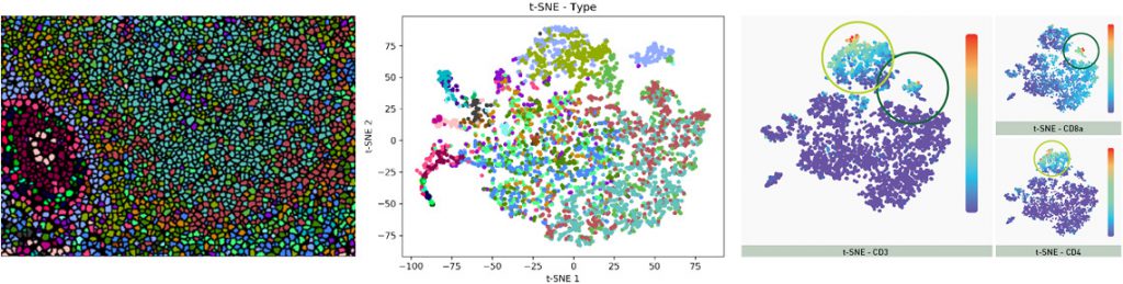
The features include automatic classification of cell populations in tissue images, automatically names phenotypes and cells to simplify the subsequent analysis. Results can be visualized in a class relationship generating a phenotypic profile and matrix, and t-SNE clustering plots for cell phenotypes.
This software will pair with Imaging Mass CytometryTM (IMC™) generated results, offering a powerful workflow to augment the analysis of highly multiplexed datasets from the Fluidigm HyperionTM Imaging System.
The PhenomapTM is an add-on module and available from now for existing and new users of the Author Module in our Visiopharm image analysis software platform (VIS).
Discover how PhenomapTM can help you – read more
About Visiopharm
Visiopharm is a world leader in Augmented Pathology™ solutions; quantitative image analysis software and Precision Pathology, solving everything from H&E to advanced fluorescence in histopathology.
Leading biopharmaceutical companies, contract research organizations (CRO), academic medical centers, and hospital diagnostic pathology labs all over the world utilize the Oncotopix® platform for tissue-based research and diagnostics.
Oncotopix® provides scientists and pathologist with a scalable software solution that fits the needs and volumes of both research and diagnostic labs.
Visit the Visiopharm APP Center visiopharm.com/appcenter, the world’s largest and fastest growing library of APPs for Diagnostic and Research. Or go directly to visiopharm.com.
Source: Visiopharm


