The DeePathology AI Challenge !

DeePathology is in a mission to bring AI to the hands of every pathologist and biological sciences researchers ! Now, we invite you to challenge our DeePathology STUDIOTM !
DeePathology has developed the DeePathology STUDIOTM – a ‘Do It Yourself’ platform that allows pathologists and researchers to build accurate AI solutions for various problems in pathology without being AI experts, super fast, and without the need to send data out of the pathology lab.
Pathologists embrace AI and exploit what digital and computational technology have to offer. We strongly believe that it is the tight collaboration between the pathology expert and the AI expert that brings pathology research and diagnostics to the next level.
The DeePathology STUDIOTM was already used by pathologists all over the world to create working solutions to a variety of problems, including Beta Amyloid Plaque detection, Astrocyte detection, Microgalia detection, Bone marrow growth plate region segmentation, Granulated/DeGranulated cells detection for skin cancer, Red blood cell detection, Ki67 cell quantification, Lung H&E wall region segmentation, Objects detection, e.g. glomerulus, Wells and Acini, H&E tumor segmentation, Region Segmentation in various IHC stains, e.g. FoxP3 and CD7, H&E tumor Infiltrating Lymphocytes region detection and quantification, H&E lymphocyte detection, H.Pylori detection and TB detection.
How did we manage to create so many AI solutions ? Our AI platform was developed to excel in finding solutions for three use cases: cell detection, object localization and classification and region segmentation. The expert pathologists teach the DeePathology STUDIOTM platform what to look for by giving positive and negative examples – providing annotations. Now, annotation sounds tedious and time consuming and we all know that pathologists do not have any spare time.
And this is why we have developed the Rapid Annotation ability where the STUDIO AI platform learns from the very first annotations what the expert is looking for and very quickly a ping pong of suggested annotations by the AI and corrections by the expert pathologist is on its way.
In addition, the STUDIO does not require huge amounts of data. We can create working solutions with only a few slides.
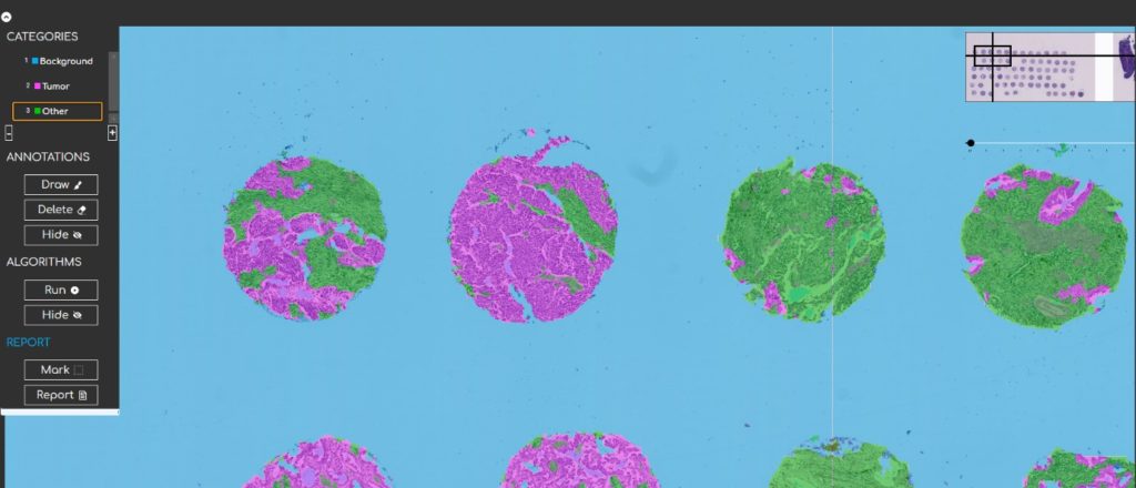
H&E TMA analysis done with the DeePathology STUDIO by Vall d’Hebron institute of Oncology.
Now, we announce the DeePathology AI Challenge !
We invite pathologists and biology sciences researchers and experts to submit a single WSI along with a specification of the problem that you would like to solve. You can send us slides digitized with any Digital Scanner or Digital Microscope. We will send a personal response to every applicant and will select three use cases to show how to create AI solutions for them in a live webinar to be held on September 15 th !
We will hold a webinar to show you how to create AI solutions in minutes – from scratch all the way to deployment on July 15th.
For more information on the DeePathology AI Challenge email: info@deepathology.ai
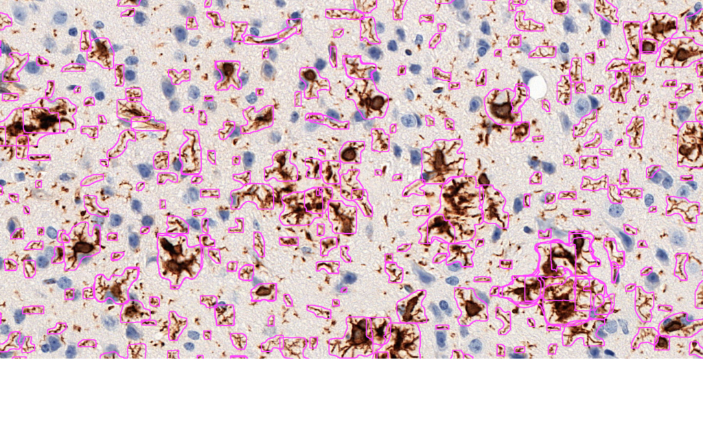
Microglia detection AI created using the DeePathology STUDIO, for Alzheimer research. Joint work with Prof. Jens Pahnke. More information can be found here: http://pahnkelab.eu/research/AI/


