PreciPoint Announces Next AI Webinar

 Computational pathology, or the use of Artificial Intelligence (AI) in the field of histopathology, holds significant potential for healthcare. For many of us, this emerging, fast-developing discipline comes with many interrogations. For pathologists, it is a change process, as they will see their working practices evolve in the coming years and decades.
At PreciPoint, we believe that communication plays a crucial part in demystifying Artificial Intelligence by bringing computer science and pathology closer together. We also believe that no pathologist must become a computer scientist in order to work with AI.
With our new series of Webinars Pathology meets Technology, we are creating a platform for both communities to learn from each other and generate more synergies.
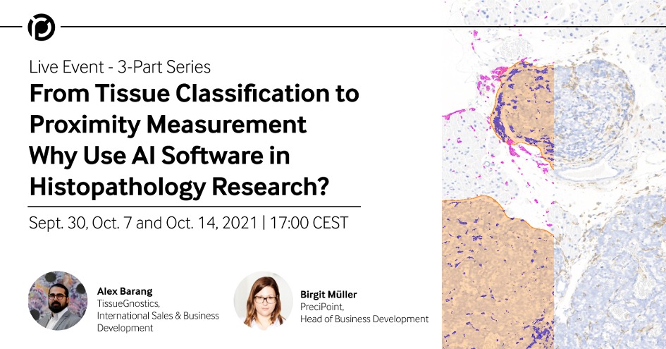
From Tissue Classification to Proximity Measurement –
Why Use AI Software in Histopathology Research
Computational Pathology • Image Analysis • AI Software
This event is part of our webinar series Pathology meets Technology.
After a short introduction on how to generate a digital image for automated analysis purposes, we will present various AI models for histopathology research applications.
The purpose of this webinar series is to explain AI software to new and upcoming AI users in the context of pathology research.
In order to do this, we will look into a spectrum of applications: From tissue classification to more advanced analyses such as nuclei segmentation and proximity measurement, you will walk away with a good understanding of what AI software can do. Along the way, we will also discuss the most compelling benefits and common challenges of using AI models that novice users should be aware of.
What will you learn?
- Why use AI in pathology research. How is it helpful for researchers?
- What is AI software and what can I use it for?
- How to use various types of AI models and for which research questions.
Part 1 – September 30, 2021 17:00 CEST / 11:00 EDT / 23:00 CST (45 min)
Overview of a classical image analysis workflow for tissue classification, and use case with machine learning for breast cancer tissue classification
Part 2 – October 7, 2021 17:00 CEST / 11:00 EDT / 23:00 CST (30 min)
Nuclei segmentation: Limitations of classical segmentation models and the use of deep learning for nuclear segmentation in breast cancer tissue.
Part 3 – October 14, 2021 17:00 CEST / 11:00 EDT / 23:00 CST (30 min)
AI powered proximity measurement analysis: Colon cancer TMA cores cell to cell interactions, and proximity areas emanating from epithelium.
Registrations:
https://us06web.zoom.us/webinar/register/1716317870981/WN_drYWvqniT7i2HpcgqhajUg
Speakers for this event:
Birgit Müller
Head of Business Development
PreciPoint GmbH
www.precipoint.com
Ms. Birgit Müller is the Head of Business Development at PreciPoint, a creator and provider of digital microscopy solutions for laboratories. In her function, Birgit is responsible for customer projects implementing digital solutions with all the changes needed both on the side of the lab as well as the technology. Ms. Müller has extensive experience with digitization projects both in laboratories as well as in industrial companies having worked in management consulting before joining PreciPoint.
Alex Barang
International Sales & Business Development
TissueGnostics
www.tissuegnostics.com
SOURCE: PreciPoint


